Echocardiography Report Template
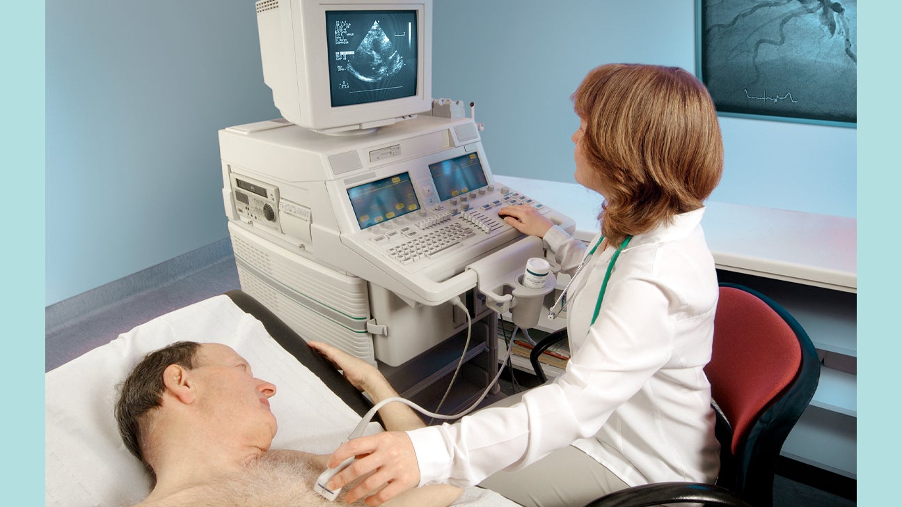
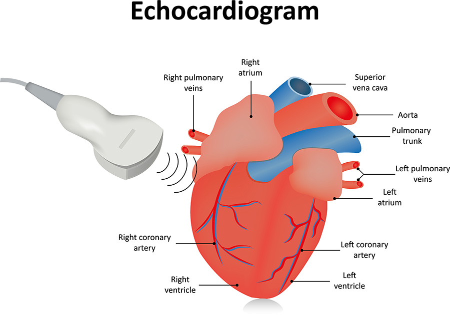
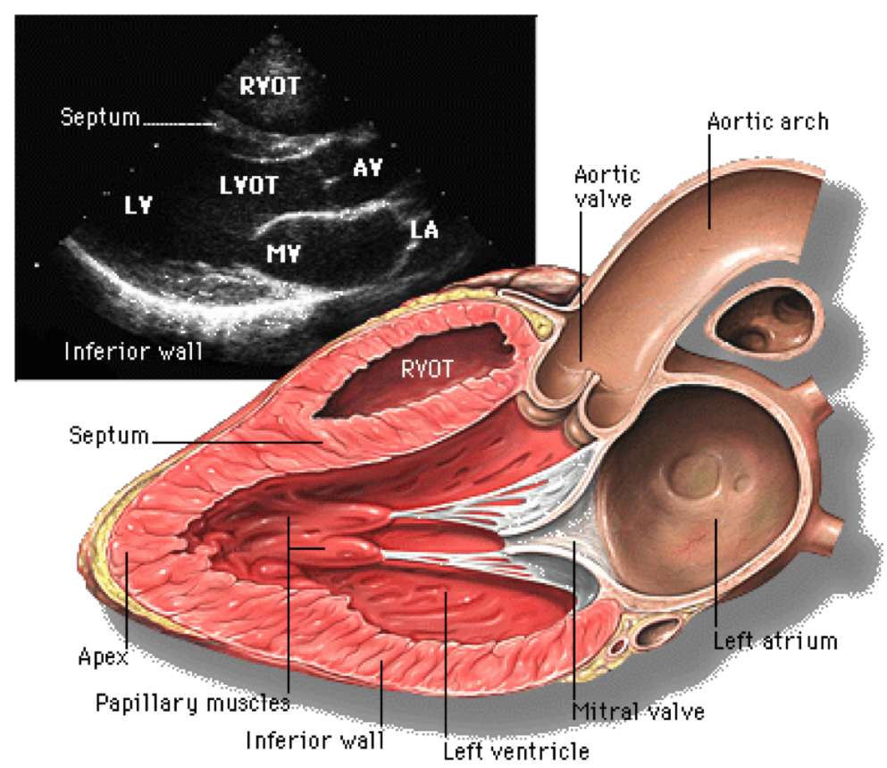
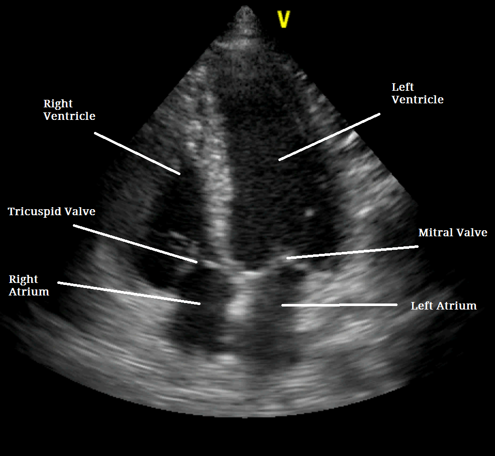
Echocardiography Report Template - It is a type of medical imaging, using standard ultrasound or doppler ultrasound. It is a noninvasive medical procedure that does not. Echocardiography uses ultrasound waves to produce an image of the heart, the heart valves, and the great vessels. During an echo, a cardiac sonographer will move a wand across this area. These images provide pictures of the heart. It allows your doctor to monitor how your heart. An ekg checks your heart's electrical activity. The image is called an echocardiogram. During an echo test, your healthcare provider uses ultrasound (high. Echocardiography, also known as cardiac ultrasound, is the use of ultrasound to examine the heart. This is done by moving an ultrasound transducer to various locations on your chest, back or abdomen in order to. It allows your doctor to monitor how your heart. Echocardiography, also known as cardiac ultrasound, is the use of ultrasound to examine the heart. An ekg checks your heart's electrical activity. Echocardiography is a test using sound waves to produce live images of your heart. These images provide pictures of the heart. There are several parts to an echocardiogram. During an echo, a cardiac sonographer will move a wand across this area. Your doctor might use this test to look at your heart’s structure and to. An echocardiogram (echo) is a graphic outline of your heart’s movement. It helps assess heart wall thickness (eg, in hypertrophy or atrophy) and. Echocardiography is a test using sound waves to produce live images of your heart. These images provide pictures of the heart. During an echo, a cardiac sonographer will move a wand across this area. An ekg checks your heart's electrical activity. Echocardiography uses ultrasound waves to produce an image of the heart, the heart valves, and the great vessels. Echocardiography uses ultrasound waves to create a picture of the heart, which is known as an echocardiogram (echo). The image is called an echocardiogram. This is done by moving an ultrasound transducer to various locations on your chest, back or abdomen in. An echocardiogram is a test that takes moving pictures of the heart with sound waves. It helps assess heart wall thickness (eg, in hypertrophy or atrophy) and. It is a type of medical imaging, using standard ultrasound or doppler ultrasound. During an echo test, your healthcare provider uses ultrasound (high. Although it has a similar name, an echocardiogram isn’t the. Although it has a similar name, an echocardiogram isn’t the same as an electrocardiogram (ecg), which is a test used to check your heart’s rhythm and electrical. During an echo test, your healthcare provider uses ultrasound (high. There are several parts to an echocardiogram. An echocardiogram is a test that takes moving pictures of the heart with sound waves. Echocardiography. During an echo, a cardiac sonographer will move a wand across this area. Your doctor might use this test to look at your heart’s structure and to. Echocardiography is the study of heart and function using sound waves. There are several parts to an echocardiogram. An echocardiogram (echo) is a graphic outline of your heart’s movement. It helps assess heart wall thickness (eg, in hypertrophy or atrophy) and. It allows your doctor to monitor how your heart. It is a type of medical imaging, using standard ultrasound or doppler ultrasound. During an echo, a cardiac sonographer will move a wand across this area. During an echo test, your healthcare provider uses ultrasound (high. Although it has a similar name, an echocardiogram isn’t the same as an electrocardiogram (ecg), which is a test used to check your heart’s rhythm and electrical. Echocardiography is a test using sound waves to produce live images of your heart. It is a noninvasive medical procedure that does not. Echocardiography is the study of heart and function using sound. These images provide pictures of the heart. An ekg checks your heart's electrical activity. Your doctor might use this test to look at your heart’s structure and to. Echocardiography is the study of heart and function using sound waves. Echocardiography is a test using sound waves to produce live images of your heart. These images provide pictures of the heart. Echocardiography is a test using sound waves to produce live images of your heart. During an echo, a cardiac sonographer will move a wand across this area. It helps assess heart wall thickness (eg, in hypertrophy or atrophy) and. There are several parts to an echocardiogram. Echocardiography uses ultrasound waves to produce an image of the heart, the heart valves, and the great vessels. An echocardiogram (echo) is a graphic outline of your heart’s movement. This is done by moving an ultrasound transducer to various locations on your chest, back or abdomen in order to. It looks for patterns to figure out if your heart is.. These images provide pictures of the heart. Echocardiography, also known as cardiac ultrasound, is the use of ultrasound to examine the heart. The image is called an echocardiogram. An echocardiogram (echo) is a graphic outline of your heart’s movement. Echocardiography is a test using sound waves to produce live images of your heart. It is a type of medical imaging, using standard ultrasound or doppler ultrasound. Echocardiography uses ultrasound waves to produce an image of the heart, the heart valves, and the great vessels. It is a noninvasive medical procedure that does not. An echocardiogram is a test that takes moving pictures of the heart with sound waves. An ekg checks your heart's electrical activity. There are several parts to an echocardiogram. Your doctor might use this test to look at your heart’s structure and to. Echocardiography is the study of heart and function using sound waves. During an echo, a cardiac sonographer will move a wand across this area. Echocardiography uses ultrasound waves to create a picture of the heart, which is known as an echocardiogram (echo). Although it has a similar name, an echocardiogram isn’t the same as an electrocardiogram (ecg), which is a test used to check your heart’s rhythm and electrical.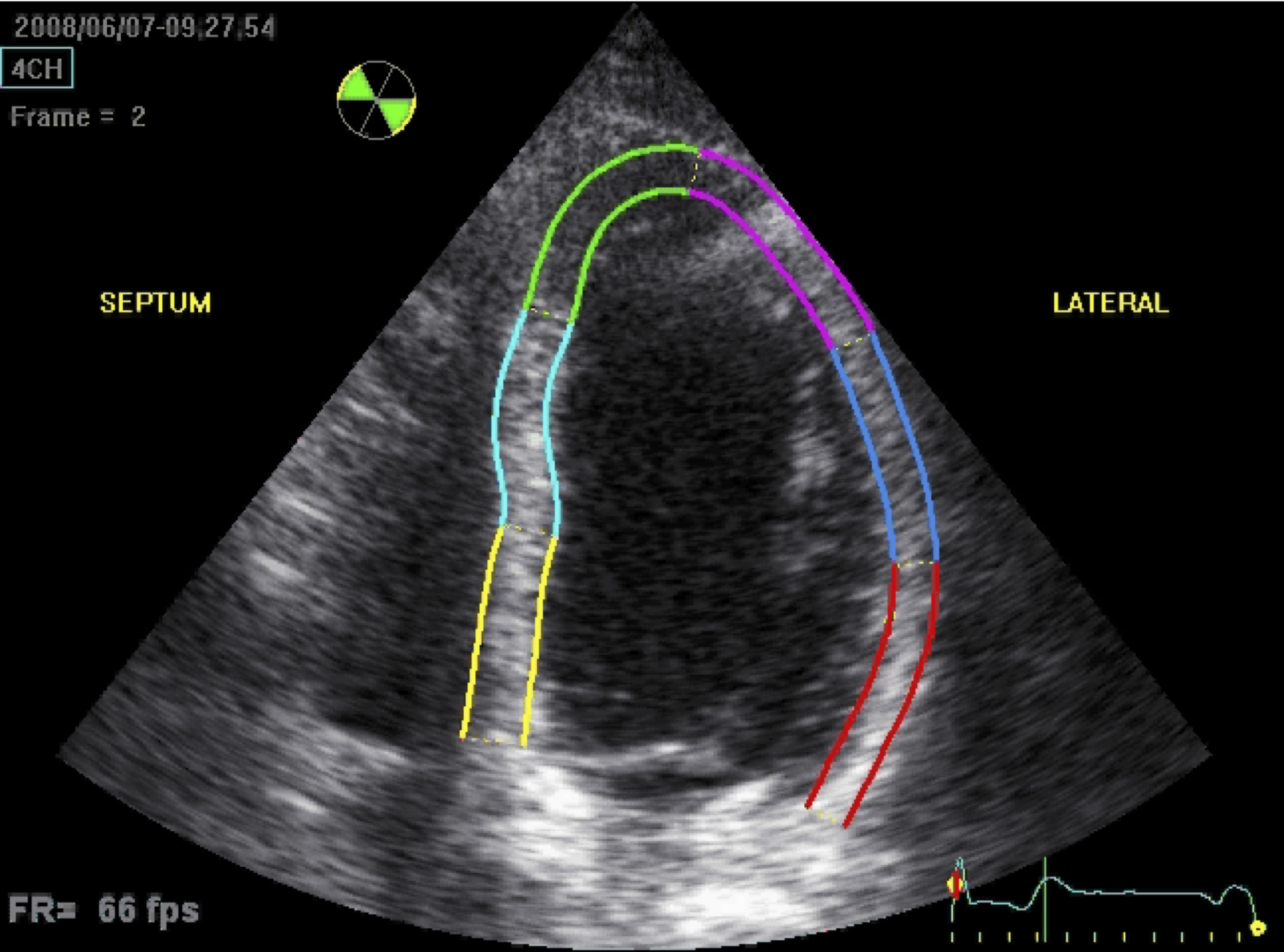
Echocardiogram Heart Ultrasound Vs
Diagnostic Imaging Center Echocardiogram at Melba Cruz blog
Echocardiogram Heart Ultrasound Vs
Echocardiography Cardiac Echo Echocardiogram Capital Area
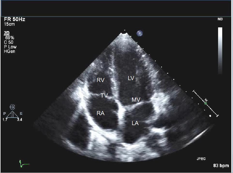
Normal Echocardiogram

Echocardiogram View Classification By Deep Learning Model Download
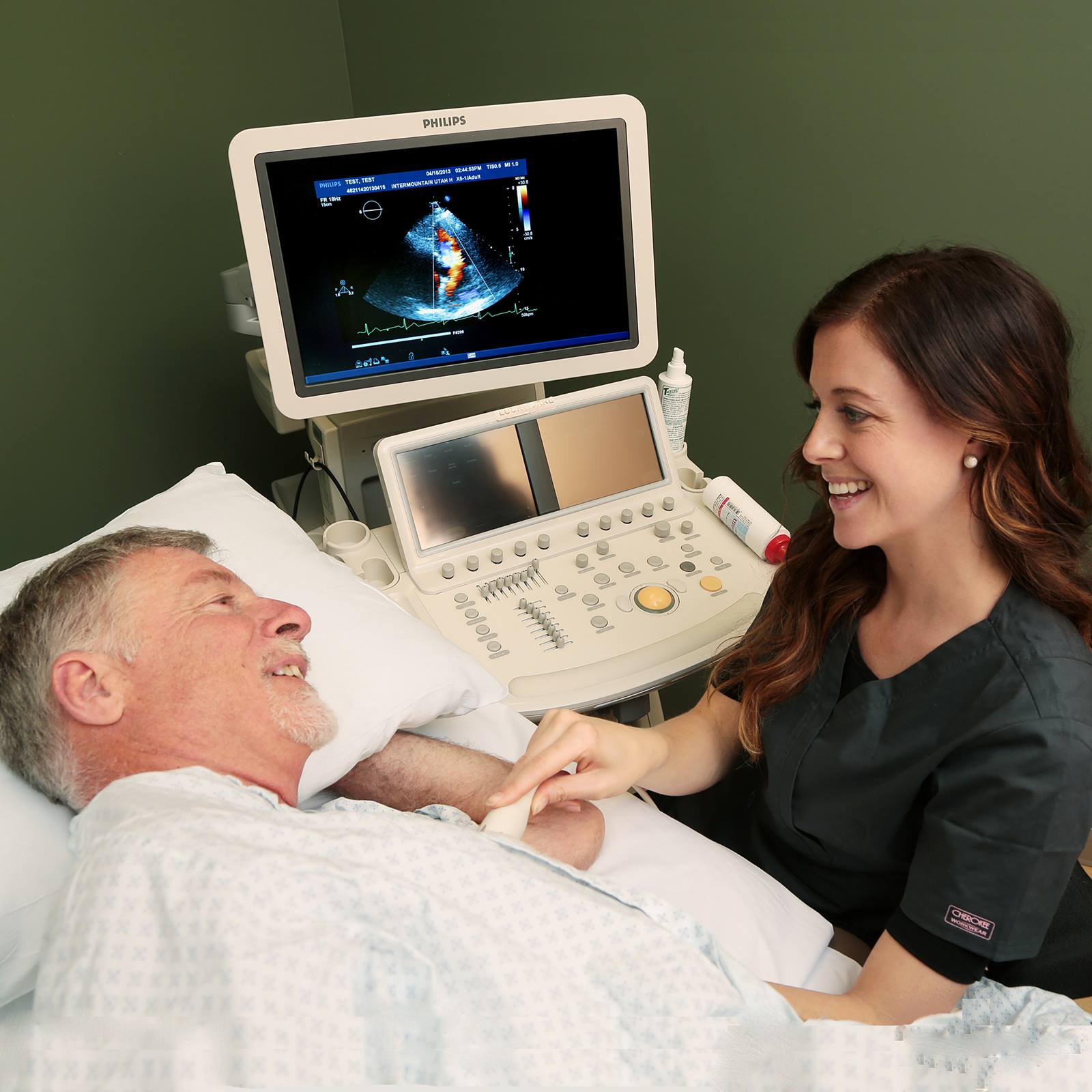
Echocardiogram What Is It, Types, Preparation, and More
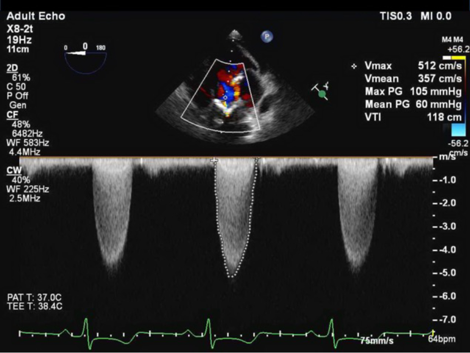
Echocardiogram Abnormal Results
Echocardiogram Report
Echocardiogram Results
It Helps Assess Heart Wall Thickness (Eg, In Hypertrophy Or Atrophy) And.
It Allows Your Doctor To Monitor How Your Heart.
This Is Done By Moving An Ultrasound Transducer To Various Locations On Your Chest, Back Or Abdomen In Order To.
It Looks For Patterns To Figure Out If Your Heart Is.
Related Post: